

The HEART score helps emergency physicians estimate the short-term risk of major adverse cardiac events (MACE) in patients presenting with chest pain and suspected acute coronary syndrome (ACS). It combines five elements – History, ECG, Age, Risk factors, and Troponin – to support decisions about further testing, observation, admission, or discharge. However, it is a risk-stratification tool, not a stand-alone test for ruling out ACS.
What is the HEART score?
Chest pain is one of the most common and challenging presentations in the emergency department (ED). The priority is to identify time-critical conditions, including myocardial infarction (MI), while avoiding unnecessary investigations and admissions in patients at low risk.
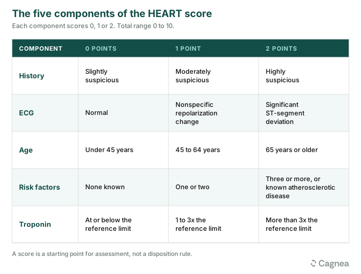
The HEART score provides a structured approach to assessing patients with suspected ACS. Each of its five components receives a score from 0 to 2, giving a total between 0 and 10.
In an original prospective validation study involving 2,440 patients, the six-week MACE rate was 1.7% among those with a HEART score of 0-3, 16.6% among those scoring 4-6, and 50.1% among those scoring 7-10. These findings demonstrate the relationship between the score and short-term risk, although event rates vary with the patient population, troponin assay, outcome definition, and clinical pathway used.
The five components of the HEART score
1. History
The history component assesses how strongly the patient’s symptoms suggest myocardial ischemia.
- 0 points: Slightly suspicious
- 1 point: Moderately suspicious
- 2 points: Highly suspicious
This is the most subjective component, and different clinicians may score the same presentation differently. Assess the character, onset, duration, radiation, exertional association, and associated symptoms rather than relying on a single feature.
Pressure-like discomfort, exertional pain, radiation to the arms or jaw, diaphoresis, and nausea can increase suspicion. Radiation to the right arm or both arms, as well as pain associated with exertion, may be more predictive of MI than radiation to the left arm alone (Tintinalli’s Emergency Medicine, Chapter 48: Chest Pain).
Do not rely exclusively on the classic presentation. Women can experience typical ischemic chest pain, but associated or less typical symptoms – including breathlessness, nausea, fatigue, and epigastric discomfort – may complicate recognition. Rare presentations such as hiccups have also been reported.
2. ECG
- 0 points: Normal ECG
- 1 point: Nonspecific repolarization abnormality
- 2 points: Significant ST-segment deviation
A normal initial ECG does not exclude ACS. Ischemic changes may evolve, so serial ECGs are important when symptoms persist, recur, or change. Compare repeat tracings with previous ECGs whenever possible.
New or dynamic ST-segment or T-wave changes can substantially alter the assessment. Patients with diagnostic ST-segment elevation or another ECG pattern requiring immediate intervention need urgent management; calculating a HEART score must not delay treatment.
3. Age
- 0 points: Younger than 45 years
- 1 point: 45-64 years
- 2 points: 65 years or older
Although MI is more common with increasing age, younger adults can also experience it, sometimes without conventional risk factors. Age should never be used alone to dismiss concerning symptoms.
4. Risk factors
The score considers hypertension, diabetes, hyperlipidemia, smoking, obesity, family history of premature coronary artery disease, and established atherosclerotic disease (e.g., prior MI, cerebrovascular accident or transient ischemic attack [CVA/TIA], and peripheral vascular disease [PVD]).
- 0 points: No known risk factors
- 1 point: One or two risk factors
- 2 points: Three or more risk factors, or established atherosclerotic disease
Take a focused risk-factor history rather than assuming that an undocumented condition is absent. Count established atherosclerotic disease as a risk factor even if other conventional risk factors are not present.
5. Troponin
In the traditional HEART score:
- 0 points: Troponin at or below the assay’s upper reference limit
- 1 point: Above the upper reference limit but no more than three times that limit
- 2 points: Greater than three times the upper reference limit
Apply the scoring criteria validated for the troponin assay in use and follow the locally approved clinical pathway. When high-sensitivity troponin is used, interpret the result and its change over time using assay-specific thresholds and validated sampling intervals.
Troponin is a marker of myocardial injury, not a specific marker of MI. It can be elevated in myocarditis or myopericarditis, acute or chronic kidney disease, pulmonary embolism, acute heart failure, sepsis, and other conditions. Interpret the result alongside the clinical presentation, ECG, and change in troponin over time.
Timing is equally important. Troponin may not have risen above the diagnostic threshold when a patient presents soon after symptom onset. An early sample can therefore be falsely reassuring, particularly with conventional assays. Follow an appropriate, validated serial troponin pathway, which may use samples at presentation and one, two, or more hours later, depending on the assay and protocol.
A normal initial result does not necessarily exclude an evolving MI. The AHA/ACC chest pain guideline recommends high-sensitivity cardiac troponin when available and emphasizes structured clinical decision pathways.
Interpreting the HEART score
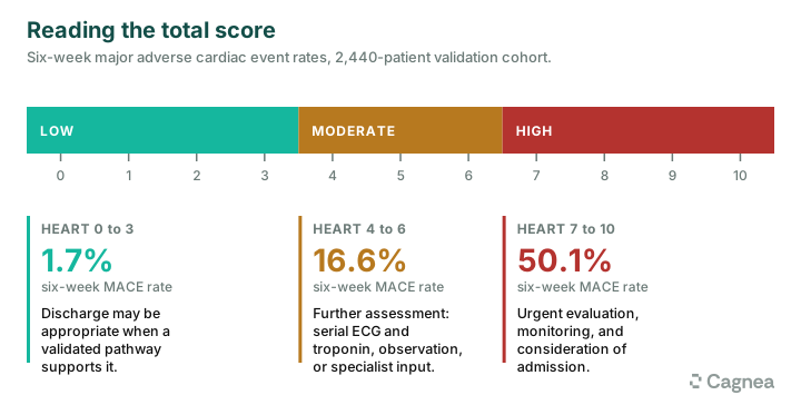
The total score ranges from 0 to 10 and is commonly divided into three risk categories. These categories help guide further evaluation but should not be used as automatic admission or discharge rules.
Low risk: 0-3
A low score may support discharge without immediate additional cardiac testing when the overall assessment and a validated clinical pathway support that decision. Consider the presenting symptoms, ECG findings, timing and results of troponin testing, alternative diagnoses, and local rule-out criteria.
Moderate risk: 4-6
These patients generally need further assessment rather than automatic discharge. Depending on the presentation and local pathway, this may include serial ECGs and troponins, observation, additional cardiac testing, or specialist discussion.
High risk: 7-10
A high score indicates substantial concern for an adverse cardiac event and generally warrants urgent evaluation, monitoring, and consideration of admission and specialist involvement. Instability, ongoing ischemic symptoms, concerning ECG changes, or a significant troponin rise should prompt immediate management rather than delay while the score is calculated.
Important limitations of the HEART score

The HEART score may underestimate risk in certain patients. Younger adults can develop MI despite having few conventional risk factors, and the score does not include every potential cause of coronary ischemia. For example, cocaine and other stimulants can cause coronary vasoconstriction, ischemia, and MI, but stimulant use is not a separate component of the score. A young patient with stimulant exposure and concerning symptoms may therefore receive a low score despite requiring further evaluation.
Interpret the score in the context of the complete clinical picture. Persistent symptoms, evolving ECG changes, or other concerning findings warrant further assessment even when the calculated score is low.
The HEART score versus the HEART Pathway
The HEART score is the 0-10 risk score based on five components.
The HEART Pathway combines the score with serial troponin testing and additional clinical criteria to identify patients who may be suitable for early discharge. The original pathway used troponin measurements at presentation and three hours, alongside a low HEART score and other criteria.
Contemporary pathways may use high-sensitivity troponin testing at different intervals, such as zero and one hour or zero and two hours. The appropriate timing depends on the assay, symptom onset, and locally validated protocol.
The HEART Pathway randomized trial found higher rates of early discharge, shorter hospital stays, and less objective cardiac testing than usual care. This supports the use of structured clinical pathways rather than the HEART score in isolation.
A practical ED example
A 48-year-old patient presents with chest discomfort that began while walking. The pain is pressure-like, but the history is not unequivocally typical of myocardial ischemia. The patient has a history of hypertension. The ECG shows nonspecific repolarization changes, and the initial troponin is within the assay’s reference range.
The HEART score is calculated as follows:
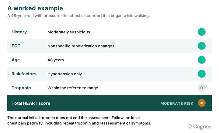
- History: Moderately suspicious – 1 point
- ECG: Nonspecific repolarization changes – 1 point
- Age: 45-64 years – 1 point
- Risk factors: One risk factor – 1 point
- Troponin: Within the reference range – 0 points
Total HEART score: 4 – moderate risk.
The normal initial troponin does not end the assessment. Further evaluation should follow the local chest pain pathway, including repeat troponin testing and reassessment of symptoms. Repeat ECGs are also indicated if symptoms persist or recur. Observation, additional testing, or admission may be appropriate depending on the evolving clinical picture.
The score helps organize the risk assessment; it does not determine disposition independently.
Documenting the decision
The medical record should make clear how the risk assessment informed the plan. Document the presenting symptoms, relevant cardiovascular risk factors, ECG interpretation, troponin results and timing, HEART score, and the clinical reasoning behind observation, admission, or discharge.
If serial testing is performed, record the results and how they affect the assessment. For discharged patients, document the follow-up plan and clear return precautions. The score should support the clinical reasoning, not replace it.
Structured documentation tools such as Cagnea may help clinicians record findings and reasoning consistently, but they should not replace clinical assessment. See Medical Decision Making Documentation in the Emergency Department.
Frequently asked questions
Can a patient with a HEART score of 3 be discharged?
Possibly. Discharge may be appropriate when the patient meets the criteria of a validated clinical decision pathway, the ECG and troponin assessment are reassuring, and no other clinical concern requires further evaluation. A score of 3 alone does not guarantee safe discharge.
Does the HEART score diagnose a heart attack?
No. It estimates the risk of short-term adverse cardiac events in patients being assessed for possible ACS. Diagnosis requires clinical evaluation, ECG interpretation, and appropriate cardiac biomarker testing.
The takeaway
The HEART score is most useful when it structures risk assessment without replacing clinical judgment. Use it alongside an appropriate troponin pathway, obtain serial ECGs when indicated, and never let a reassuring number overrule concerning clinical findings.


