

In trauma, identifying internal bleeding early can significantly influence patient management. The focused assessment with sonography in trauma (FAST) exam provides a rapid bedside assessment for free fluid in the peritoneal and pericardial spaces, without transporting an unstable patient away from the emergency department (ED).
FAST is not a comprehensive assessment of abdominal injury. It primarily detects free fluid rather than identifying the injured organ or excluding all significant injuries. A negative scan must be interpreted alongside the mechanism of injury, hemodynamic status, examination findings and clinical course.
What is the FAST exam, and when should it be performed?
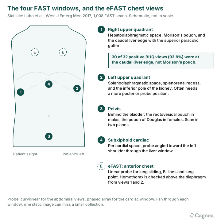
FAST stands for Focused Assessment with Sonography in Trauma. It is a point-of-care ultrasound examination designed to identify free fluid in four anatomical regions:
- Right upper quadrant (RUQ)
- Left upper quadrant (LUQ)
- Pelvis
- Pericardial space
In trauma, free fluid is presumed to represent blood until proven otherwise, particularly in patients with suspected blunt or penetrating abdominal injury. FAST is most useful when internal bleeding is suspected, especially in hemodynamically unstable patients or those with concerning findings. It forms part of the primary survey, particularly the circulation assessment, and should not delay resuscitation or definitive intervention.
A positive FAST in an unstable patient with suspected intra-abdominal hemorrhage can support an immediate decision for operative management. In a stable patient, it generally warrants further evaluation, usually with contrast-enhanced CT (CECT) of the abdomen and pelvis.
The four views of the FAST exam
A systematic examination evaluates four anatomical windows. A curvilinear probe is commonly used for the abdominal views, while a phased-array probe is useful for the cardiac window.
Four basic probe movements help ensure adequate image acquisition:
- Sliding: Moving the probe across the skin to examine adjacent areas or locate a better acoustic window.
- Rocking: Tilting the probe around its contact point to improve the view without significantly changing its position.
- Fanning: Angling the probe through different imaging planes to assess the entire anatomical space.
- Sweeping: Moving the probe continuously across a broader area to examine the full extent of a structure or space.
A single static image may miss a small collection outside the initial imaging plane. In the upper abdominal views, use an intercostal window where necessary, angling or insinuating the probe between the ribs to minimize rib shadowing.
Right upper quadrant (RUQ)
Place the probe along the right flank, usually in the mid-axillary line between the lower intercostal spaces. The liver provides an excellent acoustic window. Assess the hepatodiaphragmatic space, hepatorenal recess (Morison's pouch), right kidney, caudal edge of the liver and superior right paracolic gutter.
Examine the liver-kidney interface, the space between the liver and diaphragm, and the inferior liver tip. Fan through the region, including its posterior and inferior aspects. Do not stop at Morison's pouch: deliberately visualize the caudal tip of the liver.
In a 2017 study in the Western Journal of Emergency Medicine, Lobo et al. evaluated 1,008 FAST examinations. The region around the caudal liver edge and superior right paracolic gutter was the most frequently positive RUQ sub-region, accounting for 30 of 32 positive RUQ views (93.8%). This finding highlights the importance of extending the examination beyond Morison's pouch; it does not represent the overall sensitivity of FAST.
Left upper quadrant (LUQ)
Obtain the view along the left posterior or mid-axillary line, generally around the lower intercostal spaces. Identify the spleen, left kidney, splenorenal recess, splenodiaphragmatic space and inferior pole of the kidney with the adjacent paracolic gutter. Free fluid may collect between the spleen and kidney or between the spleen and diaphragm.
The LUQ can be challenging because of the smaller acoustic window, overlying stomach and bowel gas, and position of the spleen. A more posterior probe position or intercostal approach may be needed; fan through the region to avoid missing small collections.
Pelvic view
Place the probe suprapubically and examine the pelvis in longitudinal and transverse orientations. The bladder serves as an acoustic window. Look for free fluid posterior to the bladder and within the dependent pelvic spaces. In males, fluid may collect in the rectovesical pouch; in females, assess the pouch of Douglas, posterior to the uterus.
A partially filled bladder improves visualization. An empty bladder, bowel gas or poor positioning can make the view difficult to interpret. Small amounts of pelvic free fluid may be physiological in women of reproductive age, so interpret findings in the clinical context.
Subxiphoid cardiac view
The subxiphoid view assesses the pericardial space, particularly with suspected cardiac injury after blunt or penetrating trauma. Place the probe immediately below the xiphoid process and angle towards the patient's left shoulder, using the liver as an acoustic window. An anechoic or hypoechoic collection surrounding the heart may represent pericardial effusion or hemopericardium.
In a trauma patient, pericardial fluid with hemodynamic instability should raise concern for cardiac tamponade. Right atrial or right ventricular chamber collapse can provide additional evidence when interpreted alongside the clinical picture.
What does free fluid look like?
Free fluid typically appears as an anechoic (black) or hypoechoic collection within a space where structures would normally be apposed. It may be seen in Morison's pouch, between the spleen and kidney or diaphragm, posterior to the bladder, or surrounding the heart. Acute blood is often anechoic, although clot and debris can produce a more complex or echogenic appearance. Assess the location and overall appearance rather than relying on echogenicity alone.
eFAST: extending the examination to the chest
The extended FAST (eFAST) adds thoracic ultrasound views to the standard examination, primarily to assess for pneumothorax and hemothorax.
Assessing for pneumothorax
Use a high-frequency linear probe over the anterior chest wall, usually beginning in the upper anterior chest. Key findings include:
- Lung sliding: Normal movement of the visceral and parietal pleura against each other.
- B-lines: Vertical artifacts arising from the pleural line, indicating pleural apposition at that location; their presence excludes pneumothorax at the point of examination.
- Lung point: The transition between areas with and without lung sliding; when identified, it is highly specific for pneumothorax.
The absence of lung sliding alone does not confirm pneumothorax. It can also occur with apnea, pleural adhesions and mainstem intubation. Interpret the findings together.
Assessing for hemothorax
Hemothorax is usually assessed alongside the RUQ and LUQ views. After obtaining the upper abdominal window, slide or angle the probe cephalad, above the diaphragm, to examine the corresponding pleural space. An anechoic or complex collection above the diaphragm may indicate pleural fluid or hemothorax. Identify the diaphragm clearly to distinguish pleural fluid from intraperitoneal fluid below it.
When should you move to a RUSH examination?
FAST and eFAST are primarily trauma-focused examinations, but not every patient presenting with hypotension has sustained an injury. The Rapid Ultrasound in Shock (RUSH) examination provides a broader bedside assessment of the cardiovascular system in undifferentiated hypotension. One practical way to organize the assessment is the HI-MAP-ED mnemonic, incorporating the Pump, Tank and Pipes approach:
- H, Heart (Pump): Assess cardiac function, pericardial effusion and signs of right ventricular strain.
- I, IVC (Tank): Assess diameter and respiratory variation as part of the evaluation of intravascular volume status.
- M, Morison's pouch: Assess for abdominal free fluid, alongside the other FAST windows.
- A, Aorta (Pipes): Assess for abdominal aortic aneurysm and aortic dissection.
- P, Pulmonary: Assess for pneumothorax and pleural fluid.
- ED, Ectopic pregnancy and DVT: Evaluate for these conditions when clinically indicated.
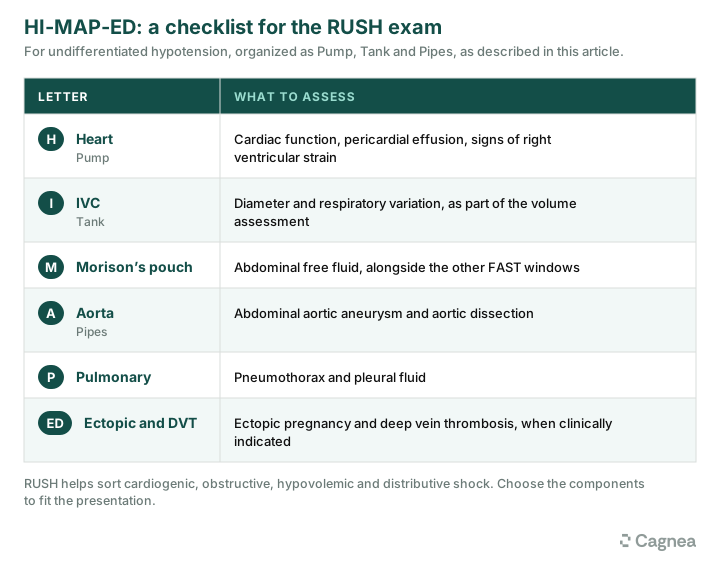
RUSH can help evaluate cardiogenic, obstructive, hypovolemic and distributive shock. Select its components according to the clinical presentation.
When does FAST miss an injury?
A negative FAST does not exclude significant intra-abdominal injury. Common causes of false-negative examinations include:
- Early bleeding: The volume of free fluid may initially be insufficient to produce a visible collection, despite significant ongoing hemorrhage.
- Retroperitoneal injuries: FAST primarily identifies intraperitoneal and pericardial fluid; it is not reliable for injuries involving the kidneys, pancreas, aorta or major retroperitoneal vessels.
- Solid organ, bowel and mesenteric injuries: Liver and splenic injuries may occur without significant hemoperitoneum. Hollow viscus and mesenteric injuries may also produce too little free fluid to be detected.
- Technical limitations: Obesity, subcutaneous emphysema, bowel gas, poor positioning, rib shadowing, inadequate probe selection and operator experience can affect image quality.
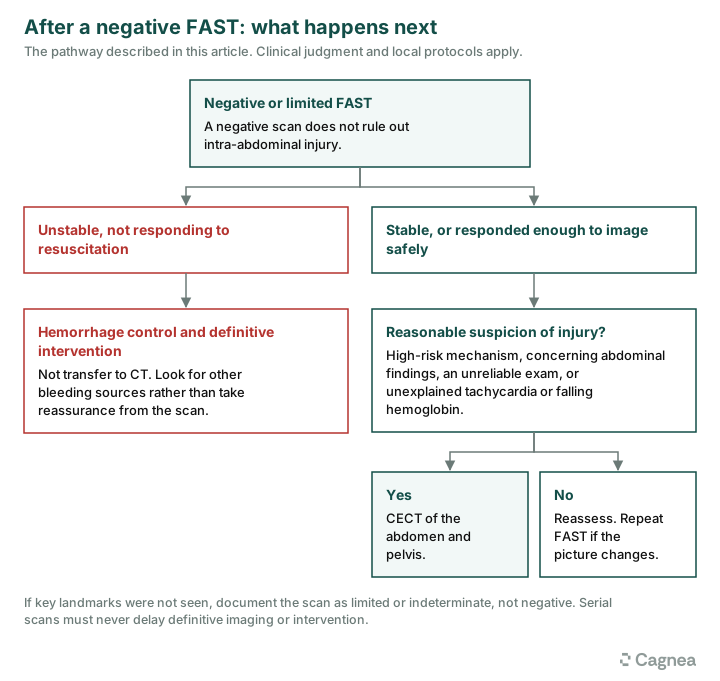
If important anatomical landmarks are not adequately visualized, document the examination as limited or indeterminate rather than negative. These limitations are addressed in the AIUM practice parameter for eFAST.
When should FAST be repeated?
FAST is a snapshot of the patient's condition. Repeat it when clinical suspicion remains high, the patient deteriorates, or the initial examination was technically limited. Examples include persistent or worsening tachycardia or hypotension, an unexplained fall in hemoglobin, increasing blood product requirements, or new abdominal tenderness or distension.
There is no universally applicable fixed interval; timing should be guided by the clinical course, with reassessment whenever the hemodynamic picture changes. Serial examinations can improve sensitivity, but must never delay definitive imaging or intervention when indicated. The role and limitations of serial examinations are discussed in this review of eFAST in trauma.
When should you proceed to CECT despite a negative eFAST?
A negative eFAST should not be the sole reason to withhold CT when significant abdominal injury is suspected. In hemodynamically stable patients, CECT of the abdomen and pelvis is the preferred investigation when there is reasonable suspicion of intra-abdominal injury. It provides a more comprehensive assessment of solid organs, hollow viscera, mesentery, retroperitoneum and vascular structures.
Consider CECT despite a negative eFAST in patients with:
- High-risk mechanism or associated injuries: Significant motor vehicle collisions, falls from height, seatbelt sign, pelvic fractures or multiple long-bone fractures.
- Concerning abdominal findings: Persistent pain or tenderness, peritoneal signs, abdominal distension or hematuria.
- Unreliable examination: Altered sensorium, intoxication, distracting injuries or inability to perform reliable serial examinations.
- Unexplained clinical or laboratory abnormalities: Persistent tachycardia, falling hemoglobin or suspicion of bowel, mesenteric, pancreatic or retroperitoneal injury.
In an unstable patient who is not responding to resuscitation, the priority is hemorrhage control and definitive intervention, not transfer to CT. A negative FAST should prompt assessment for other bleeding sources rather than reassurance. CT is appropriate when the patient is stable or has responded sufficiently to resuscitation to undergo imaging safely.
A negative FAST does not rule out intra-abdominal injury. In hemodynamically stable patients, CECT remains the preferred imaging modality when injury is suspected.
Documenting the FAST examination
Document the indication, views obtained and adequacy of visualization. Record the presence or absence of free fluid in the RUQ, LUQ, pelvis and pericardial space, specifically noting Morison's pouch and the caudal liver edge. For eFAST, include the lung and pleural findings. State technical limitations, the overall interpretation and the clinical plan; do not document the scan simply as "FAST negative" if views were inadequate.
Structured POCUS documentation can improve consistency and communication. In Cagnea's article, POCUS in the ED Meets AI Interpretation, the potential roles of AI include supporting image acquisition, quantifying findings and flagging abnormalities. In FAST, these tools may help operators obtain adequate views and identify possible free fluid. However, interpretation still depends on clinical context and physician judgment. AI should complement, not replace, the clinician's assessment.
The takeaway
FAST is a valuable bedside tool for rapidly identifying free fluid during the trauma primary survey. Systematic acquisition, adequate visualization of all four windows and awareness of its limitations are essential.
eFAST extends the assessment to the chest, while RUSH provides a broader approach to undifferentiated shock. A negative FAST does not rule out intra-abdominal injury: repeat the scan when the clinical picture changes and proceed to CECT when the mechanism, examination or clinical course warrants it. The value of FAST lies not just in identifying what is present, but in recognizing what the examination may have missed.
Where Cagnea fits
Cagnea drafts the encounter note from the conversation, so the clinician reviews and signs rather than types. The scan findings, the reasoning and the decision about CT stay with the physician. You can see how it works, read about MDM documentation in the emergency department, or check what it costs.


